


Advanced Retinal Imaging vs. Standard Dilation: How Optomap Technology Catches Hidden Eye Health Risks Early
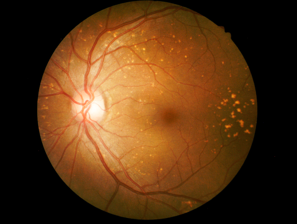
Advanced Retinal Imaging vs. Standard Dilation: How Optomap Technology Catches Hidden Eye Health Risks Early
Many eye diseases can begin without pain, blurry vision, or other noticeable symptoms. By the time vision changes occur, damage may already be present. Advanced retinal imaging helps eye doctors examine the back of the eye and identify potential concerns before they begin affecting everyday vision.
At Bogey Hills Vision Center in Saint Charles, Optomap retinal imaging is available as part of the practice’s advanced eye care technology. It provides detailed wide-field images that help doctors evaluate and document retinal health.
What Happens During a Dilated Eye Exam?
During dilation, special eye drops widen the pupils so the eye doctor can examine the retina, optic nerve, and blood vessels. This remains an important part of comprehensive eye care, particularly for patients with symptoms, existing eye conditions, or elevated risk factors.
However, dilation can temporarily cause light sensitivity and blurred near vision. Patients may need sunglasses afterward and may have difficulty reading, working on a computer, or driving until the effects wear off.
How Optomap Retinal Imaging Works
Optomap uses ultra-widefield retinal imaging to create a digital image of the retina. The technology can capture up to 200 degrees of the retina in a single image, including peripheral areas that may be difficult to document with traditional retinal photography.
The process is quick, noninvasive, and typically does not require dilating drops. The images are available immediately, allowing the eye doctor to review the retina with the patient and explain any areas that require attention.
Eye Health Risks Optomap May Help Detect
A detailed view of the retina can help identify changes associated with conditions such as:
• Diabetic retinopathy and abnormal retinal blood vessels
• Retinal holes, tears, or signs of detachment
• Age-related macular degeneration
• Changes involving the optic nerve or peripheral retina
• Other abnormalities that may not yet cause symptoms
At Bogey Hills Vision Center, we use Optos Monaco technology to help evaluate retinal conditions, including diabetic retinopathy, macular degeneration, retinal holes, and retinal detachments.
Optomap vs. Dilation
Optomap and dilation provide different types of information. Optomap creates a permanent digital record that can be stored and compared during future eye exams. This makes it easier to identify subtle retinal changes over time.
Dilation allows the eye doctor to examine the inside of the eye directly and may still be recommended based on symptoms, medical history, age, or findings from retinal imaging. Optomap should not be viewed as a replacement for dilation in every situation. Instead, the two methods can complement each other and support a more complete evaluation.
Schedule Your Visit With Bogey Hills Vision Center
Routine retinal imaging is valuable even when vision seems clear. Conditions such as diabetic retinopathy, macular degeneration, and certain retinal problems can develop quietly. Establishing a retinal health baseline gives our Saint Charles eye doctor an important reference point for future visits.
Schedule an eye exam with Bogey Hills Vision Center to discover how Optomap retinal imaging can support early detection and protect your eye health. Visit our office in Saint Charles, Missouri, or call (636) 946-1176 to book an appointment today.


Office Hours
- Monday 8:30am - 5:00pm
- Tuesday 8:30am - 5:00pm
- Wednesday 8:30am - 5:00pm
- Thursday 11:00am - 6:30pm
- Friday 8:30am - 5:00pm
- Saturday Closed
- Sunday Closed
© 2026 Bogey Hills Vision Center | All rights Reserved | Accessibility Statement | Privacy Policy | Terms and Conditions | Sitemap
Powered by:
